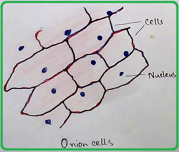
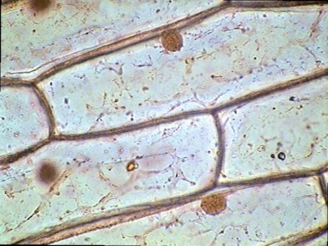
43 onion cells under microscope with labels
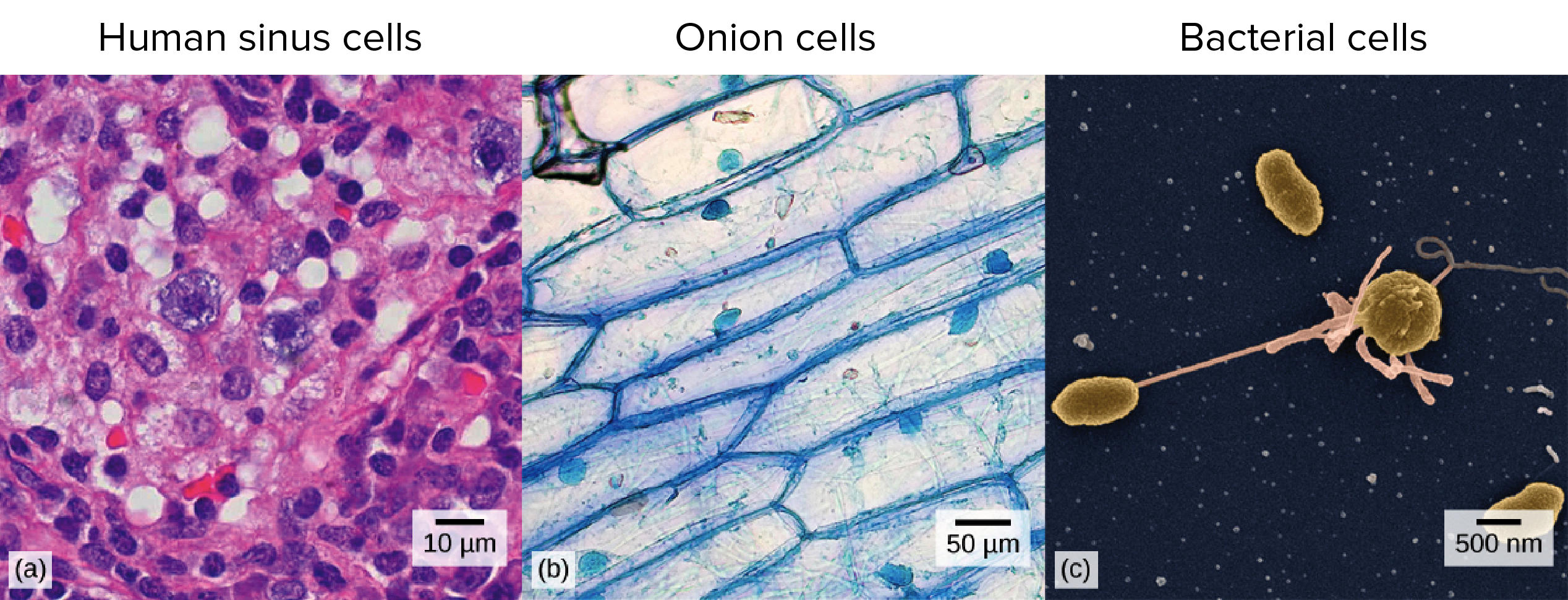
Looking at the Structure of Cells in the Microscope A typical animal cell is 10-20 μm in diameter, which is about one-fifth the size of the smallest particle visible to the naked eye. It was not until good light microscopes became available in the early part of the nineteenth century that all plant and animal tissues were discovered to be aggregates of individual cells. This discovery, proposed as the cell doctrine by Schleiden and Schwann ... DOC The Onion Cell Lab - chsd.us Place the single layer of onion cell epithelium on a glass slide. Make sure that you do not fold it over or wrinkle it. Place a drop of iodine stain on your onion tissue. Put the cover slip on the stained tissue and gently tap out any air bubbles. Observe the cells under 4x, 10x, and 40x with the diaphragm wide open.
VIEWING PLANT CELLS UNDER THE MICROSCOPE: onion ... MICROSCOPE: onion cell preparation. This method allows students to view plant cells under the microscope. A single layer of onion.
Onion cells under microscope with labels
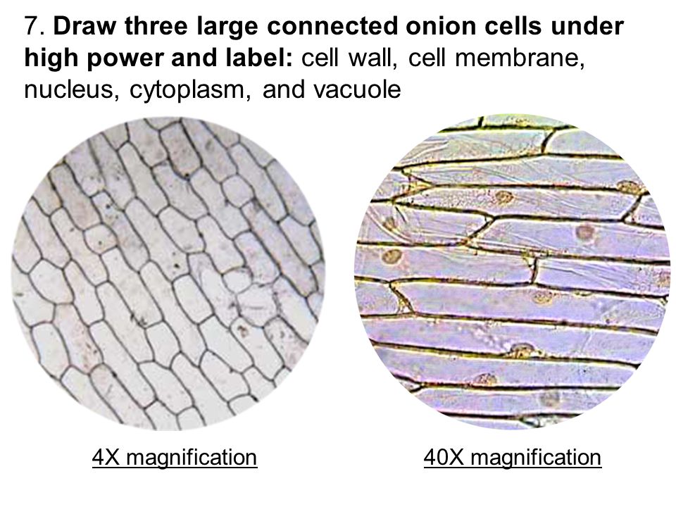
PDF Onion Cells - Investigation 1. Observe the onion tissue under the microscope at 4x, 10x and 40x with lots of light (open diaphragm). 2. Then slowly close the diaphragm while observing the image to find the best light for seeing cellular details. 3. Draw a section of onion skin cells at 10x magnification. 4. Switch to 40x and draw one cell and label it. PDF Onion Cell Lab Research Biology Onion Cell Lab page 1 of 3 Onion Cell Lab After you have completed the rest of this lab come back to this cover page DRAW & LABEL AN ONION CELL WITH ALL THE PARTS / ORGANELLES YOU OBSERVE UNDER 40X. Purpose: To observe and identify major plant cell structures and to relate the structure of the cell to its function. Materials: 1 ... Onion Skin Cells - Investigation 5. Observe the onion tissue under the microscope at 4x, 10x and 40x with lots of light (open diaphragm). Then slowly close the diaphragm while observing the image to find the best light for seeing cellular details. 6. Draw a section of onion skin cells at 10x magnification. Then switch to 40x and draw one cell and label it.
Onion cells under microscope with labels. How to observe cells under a microscope - BBC Bitesize All living organisms are made up of cells. Cells are the smallest part of a living organism and are around 0.01 mm - 0.03 mm long. To look at a cell close up a microscope needs to be used. Observing Cork Cells Under The Microscope » Microscope Club Place the cork dust on the microscope slide with a drop of water, then add another water droplet on top of the cork sample. Cover the prepared slide with a cover slip. Method 2 Alternatively, slice thin cork slices, making sure that ample light can pass through the slice, allowing you to see the cell layout and the individual cells. Observing Onion Cells Under The Microscope » Microscope Club Afterwards, carefully mount the prepared and stained onion cell slide onto the microscope stage. Make sure that the cover slip is perfectly aligned with the microscope slide, and that any excess stain has been wiped off. Secure the slide on the stage using the stage clips. Onion Cell Lab Report.docx - Onion Cell Lab Report By Onion Cell Lab Report By : Nawaf Almalki Introduction: Many things that are viewed using a microscope, particularly cells, can appear quite transparent under the microscope. The internal parts of the cells, the organelles, are so transparent that they are often difficult to see. Biologists have developed a number of stains that help them see the cells and their organelles by adding color to ...
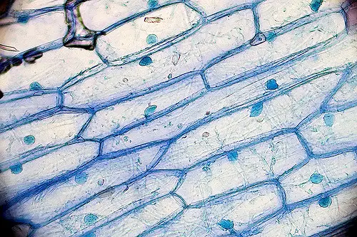
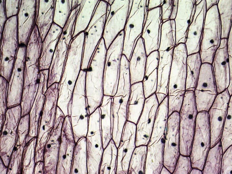
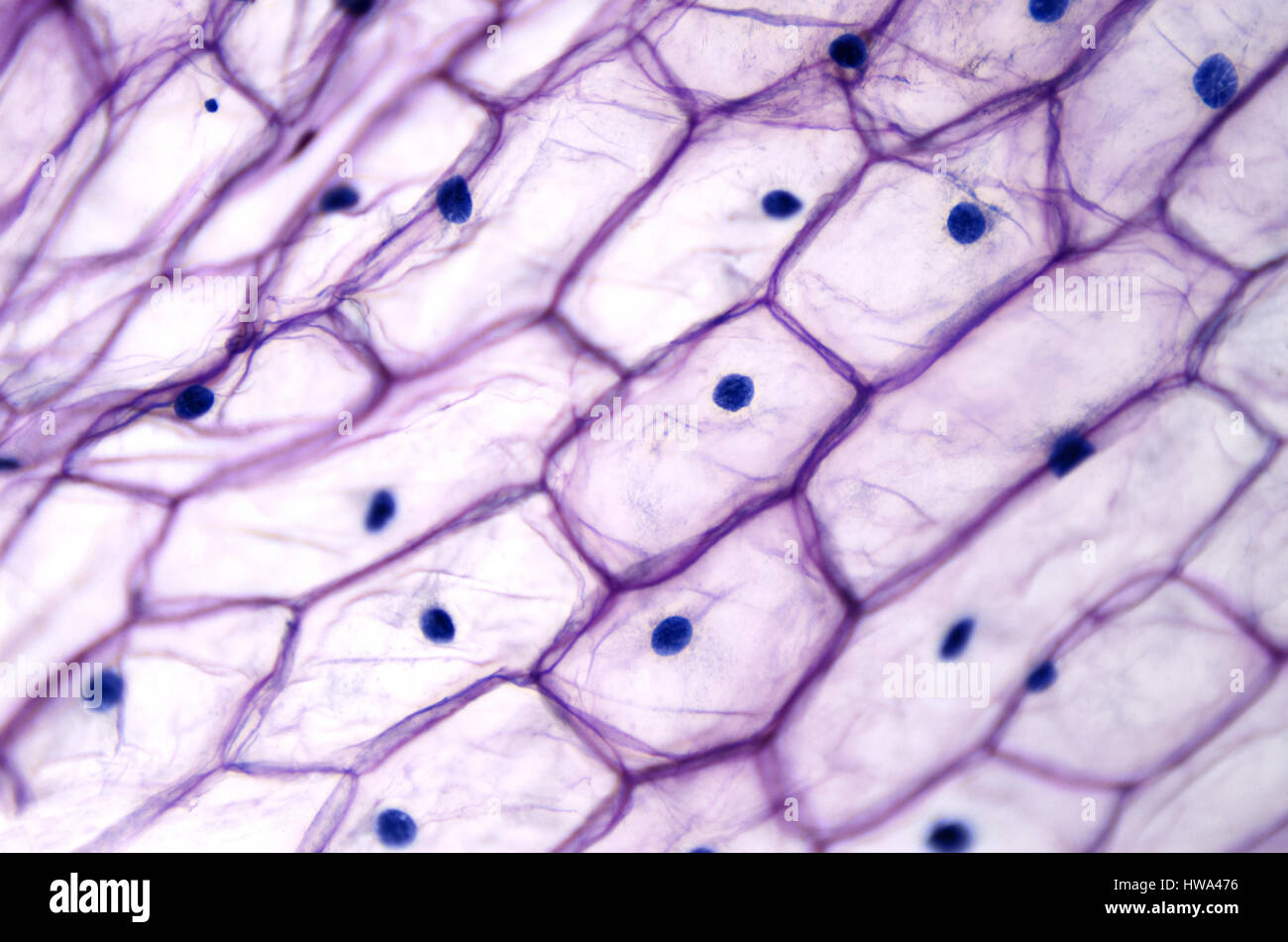
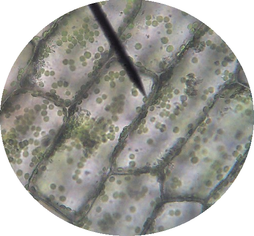
Epidermal onion cells under a microscope. Plant cells appear polygonal ... Observing onion cells under the microscope. For this microscope experiment, the thin membrane will be used to observe the cells. An easy beginner experiment. Jessica Williams. Ideas for Work. Scanning Electron Micrograph. Microscopy. Cell Division. Animal Cell. Macro And Micro. Petri Dish. Plant Cell Under Microscope 40X / Plant Cells Under ... - Blogger A number and title (ex: There four focus level in compound microscope 4x,10x,40x and 100x just place your prepared slide of plant between light and slide stand and focus on 40x or 100x you can easily see plant cells under microscope. In this simple microscope experiment, we will compare plant cells and animal cells. DOC Plant and Animal Cells Microscope Lab - hillsboro.k12.oh.us Make a drawing of one onion cell, labeling all of its parts as you observe them. (At minimum you should observe the nucleus, cell wall, and cytoplasm.) Cheek cells 1. To view cheek cells, gently scrape the inside lining of your cheek with a toothpick. DO NOT GOUGE THE INSIDE OF YOUR CHEEK! (We will observe blood cells in a future lab!!) 2. How to Observe Onion Cells under a Microscope 19 Dec 2015 — Learn how to prepare an onion for observation in order to observe the individual cells under a microscope. Staining cells included!
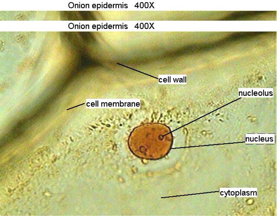
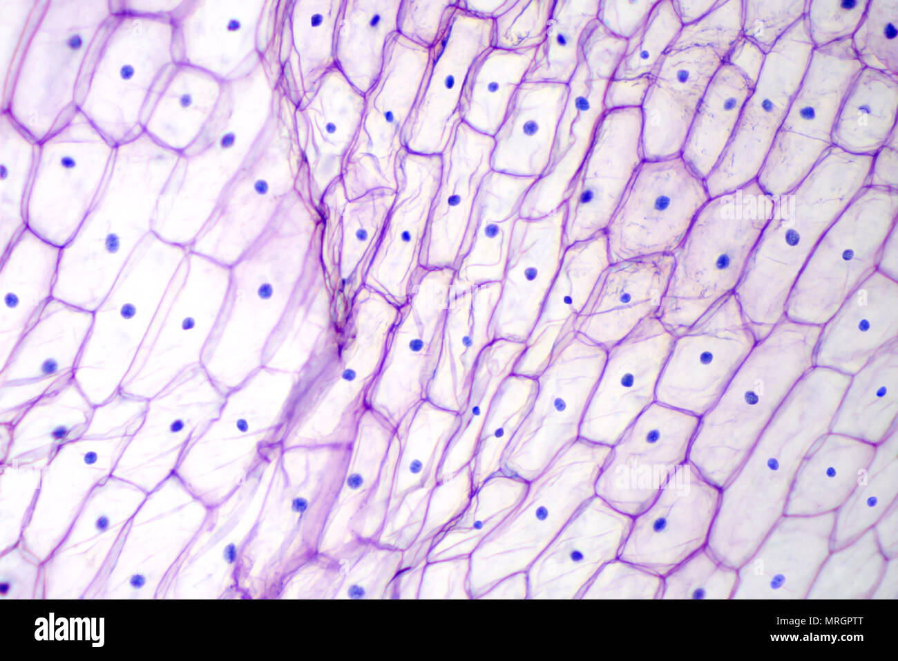
Cheek Cells Under a Microscope - Requirements/Preparation/Staining smear the cotton swab on to the center (part containing the saline drop) of the clean slide for about 4 seconds to get the cells on to the center of the slide add a drop of methylene blue solution on to the smear and gently place a cover slip on top (to cover the stain and the cells) Onion Peels Observed Under the Microscope | Confirmation Point Onion Peels Observed Under the Microscope Cells present in onion peel can be observed under microscope. For this onion peels are first isolated. For this experiment outer most scale of the onion is removed and is cut into four equal halves. It is a monocot plant. Then with the help of a pairs of forcep the scale of onion is peeled out. Onion Epidermis - kuensting.org Onion epidermal cells, iodine stain, 400X. The nucleus of an onion epidermal cell, 1000X magnification. ... Plant Cell Under Microscope 100X Labeled : Integrated Role Of Ros And ... A picture showing a microscope an onion and glass slides. At 400x, micrograph width = 0.2 mm; View and focus specimens under a microscope. • handouts with labeled animal cell. A student is observing onion cells under the microscope with a 100x magnification. • print the powerpoint presentation: • handouts with labeled plant cell.
Plasmolized red onion cells under microscope : r/biology
Onion cells under the microscope: 40X - 100X - 400X - YouTube under the #microscope: 40X - 100X - 400X

Onion Epidermis
Viewing onion cells under a light microscope in biology - Shutterstock Find Viewing Onion Cells Under Light Microscope stock images in HD and millions of other royalty-free stock photos, illustrations and vectors in the Shutterstock collection. Thousands of new, high-quality pictures added every day.

How to Observe Onion Cells under a Microscope - Blog, She Wrote
Animal Cell Mitosis Under Microscope - Casey Sillman The division of the cell in two (cytokinesis) occurs chromosomes decondense (no longer visible under light microscope). In cell biology, mitosis (/maɪˈtoʊsɪs/) is a part of the cell cycle in which replicated chromosomes are separated into two new nuclei. Plant cells do not have centrioles like animal cells, just centrosomes.
Onion Cell Under Microscope 40X Stock Image - Image of cell ...
Onion Plant Cell Under Microscope Labeled - Ismael Dauila Explore diffusion/osmosis by looking at onion cells under the microscope. It is used for treating a parasite disease called ich (ichthyophthirius multifiliis; Label the cell wall and chloroplasts. Students will observe plant cells using a light microscope.
Solved] Fig. 2.7. Stained cells of fleshy onion (Allium cepa ...
Plant Cell Under Microscope 40X Labeled - Blogger The bulb of an onion is formed from modified leaves. Put a drop of water on the microscope slides. Unlike most plant cells, this species do not have a cell wall. See how a generalized structure of an animal cell and plant cell look with labeled diagrams. Under the microscope, plant cells are seen as large rectangular interlocking blocks.
Cells Under A Microscope by Jaimarie Nelson
Plant Cell Under Microscope Observation : Grass cells under a ... Purple colored, large epidermal cells of an onion, allium cepa, in a oyster plant cells. Your plant cells under microscope stock images are ready. Observe the slide under microscope. Observe the labeled diagram of plant cell. Draw the structure that you see under microscope!! Place the glass slide onto the stage.

Onion Cell.pdf - Name: Junior Karimatsenga The Onion Cell 1 ...
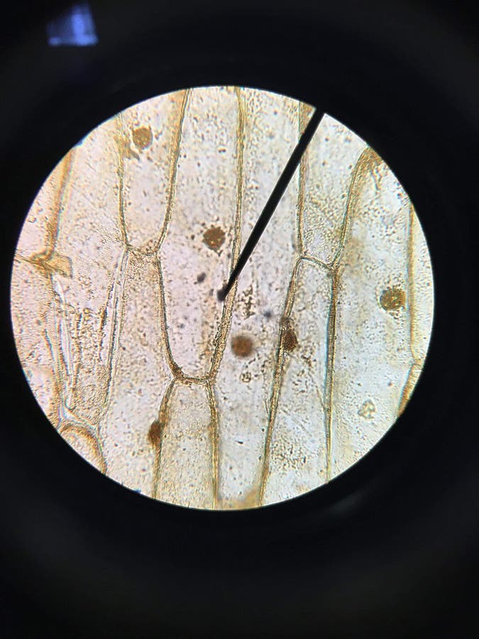
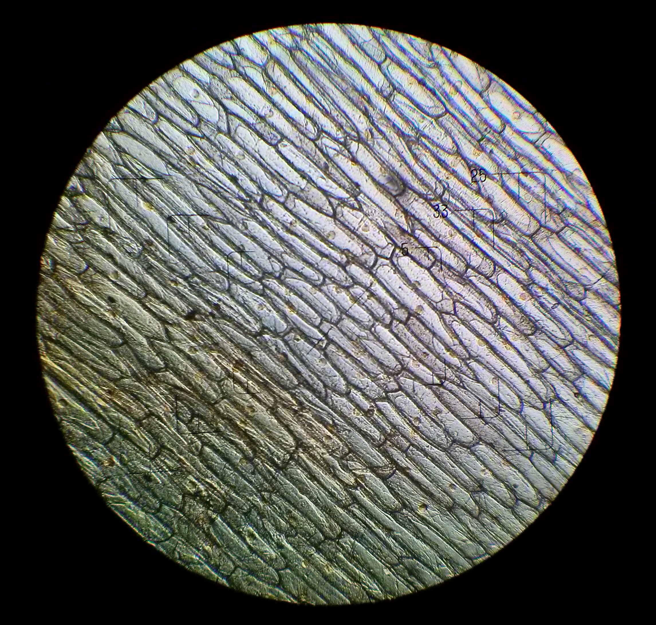
Hypothesis onion cell will be visible when viewed in - Course Hero 12.Again, looking from the SIDE of the microscope, rotate the lenses to the highest-powered lens (40x objective). If you need to, use the fine focus knob (the smallest knob) to get the image into focus. You should see a dark blob in the middle of each cell. 13.In your lab notebook, draw a picture of what you see. Label the picture "Onion skin cells 400x".
Onion Cells Under a Microscope - Requirements/Preparation ...
Onion Cells Under a Microscope - Requirements/Preparation ... Add a drop of iodine solution on the onion membrane (or methylene blue) Gently lay a microscopic cover slip on the membrane and press it down gently using a needle to remove air bubbles. Touch a blotting paper on one side of the slide to drain excess iodine/water solution, Place the slide on the microscope stage under low power to observe.
Onion Peels Observed Under the Microscope | Confirmation Point
Onion Cells Under a Microscope (100x-2500x) - YouTube 567 views Feb 24, 2021 In this video you will see onion cells under a microscope (100x-2500x) as is, without any coloring. To observe the onion cells the thin membrane is used. It can easil ...more...
322 Onion Cells Photos - Free & Royalty-Free Stock Photos ...
Plant Cell Under Microscope Labeled 40X - Sadie Bermingham Cells and viewing them under the microscope. A small square of a red onion skin (membrane) was observed under a microscope at high power (x40) magnification. (iv) describe how you applied the stain. They must draw and label the nucleus, cell membrane set up your microscope, place the onion root slide on the stage and focus on low (40x) power.

Onion Epidermis
Onion Cell Under Microscope Labeled a scientist is observing onion cells and human cheek cells under a microscope label the cell wall, nucleus, and cytoplasm draw and label your observations: label the nucleus, cell wall, and cytoplasm of one onion cell students know the characteristics that distinguish plant cells from animal cells, including chloroplasts and cell walls once you …
Onion cells hi-res stock photography and images - Alamy
Onion Skin Cells - Investigation 5. Observe the onion tissue under the microscope at 4x, 10x and 40x with lots of light (open diaphragm). Then slowly close the diaphragm while observing the image to find the best light for seeing cellular details. 6. Draw a section of onion skin cells at 10x magnification. Then switch to 40x and draw one cell and label it.
File:Onion cells under the light microscope.jpg - Wikimedia ...
PDF Onion Cell Lab Research Biology Onion Cell Lab page 1 of 3 Onion Cell Lab After you have completed the rest of this lab come back to this cover page DRAW & LABEL AN ONION CELL WITH ALL THE PARTS / ORGANELLES YOU OBSERVE UNDER 40X. Purpose: To observe and identify major plant cell structures and to relate the structure of the cell to its function. Materials: 1 ...
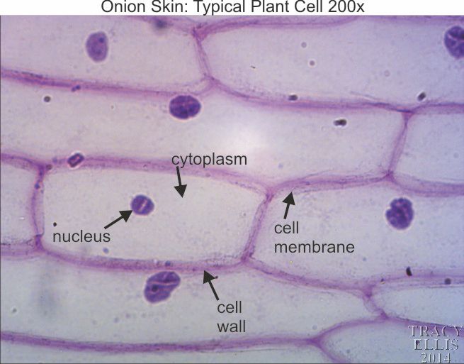
Onion skin 200x - Dissection Connection
PDF Onion Cells - Investigation 1. Observe the onion tissue under the microscope at 4x, 10x and 40x with lots of light (open diaphragm). 2. Then slowly close the diaphragm while observing the image to find the best light for seeing cellular details. 3. Draw a section of onion skin cells at 10x magnification. 4. Switch to 40x and draw one cell and label it.
Biology Pictures: Onion Cells under Microscope

Onion Skin Epidermis Sample under microscope 4x,10x Magnification

onion cells - Google Images | Ilustrações, Felipão, Estampas

Onion cells under microscopes | News | Wimbledon High School
Cells - Cells to systems - KS3 Biology Revision - BBC Bitesize

Lab #1 microscope structure & function

File:Onion skin cells under a microscope.jpg - Wikimedia Commons

Sketch the onion peel cell as seen under the microscope ...
OSMOSIS IN ONION CELLS

Lab Manual Exercise # 1
Lab: Comparing Plant and Animal Cells - ppt video online download
Onion Cell prep

Onion Cells under Microscope
Plant Cell Lab (Makeup)

How can we observe the onion peel cell
166,424 Cell wall Images, Stock Photos & Vectors | Shutterstock

Microscope Lc - Lessons - Blendspace
Onion cells under the microscope under two CONDITIONS ...
Onion epidermis under light microscope. Purple colored, large ...

Under The Microscope Onion Cells Stock Photo, Picture And ...

Microscope Cell Lab: Cheek, Onion, Zebrina | SchoolWorkHelper

TrentCollege Biology on Twitter: "Onion cells under the ...
Required Practical Review
Biology

Onion Epidermis
Intro to cells (article) | Khan Academy

Onion epidermis with large cells under microscope art print poster

Cell structure Learning Intention: - ppt video online download

Of the given slide observations spot the correct sequence ...
Post a Comment for "43 onion cells under microscope with labels"