40 brain mri with labels
Atlas of BRAIN MRI - W-Radiology Brain magnetic resonance imaging (MRI) is a common medical imaging method that allows clinicians to examine the brain's anatomy (1). It uses a magnetic field and radio waves to produce detailed images of the brain and the brainstem to detect various conditions (2). Labeled imaging anatomy cases | Radiology Reference Article ... This article lists a series of labeled imaging anatomy cases by body region and modality. Brain CT head: non-contrast axial CT head: non-contrast coronal CT head: non-contrast sagittal CT head: angiogram axial CT head: angiogram coronal CT...
Labeling Brain Structures - John Muschelli 1 Labels in template space. In Processing Within-Visit MRI, we registered the T1 image to the Eve template using a non-linear registration (SyN) (Avants et al. 2008). Also, we applied this transformation to the intensity-normalized T1, T2, and FLAIR images, so that these image are located in the same space as the Eve atlases. We can overlay the ...
Brain mri with labels
Learning-based 3T brain MRI segmentation with guidance from 7T MRI labeling Abstract Purpose: Segmentation of brain magnetic resonance (MR) images into white matter (WM), gray matter (GM), and cerebrospinal fluid (CSF) is crucial for brain structural measurement and disease diagnosis. Learning-based segmentation methods depend largely on the availability of good training ground truth. The MRI Dataset with labels. | Download Scientific Diagram Download scientific diagram | The MRI Dataset with labels. from publication: Detection of brain abnormality by a novel Lu-Net deep neural CNN model from MR images | The identification and ... Brain MRI: How to read MRI brain scan | Kenhub MRI is the most sensitive imaging method when it comes to examining the structure of the brain and spinal cord. It works by exciting the tissue hydrogen protons, which in turn emit electromagnetic signals back to the MRI machine. The MRI machine detects their intensity and translates it into a gray-scale MRI image.
Brain mri with labels. CaseStacks.com - MRI Brain Anatomy Labelled radiographs and CT/MRI series teaching anatomy with a level of detail appropriate for medical students and junior residents. Pelvis Pelvic MRI anatomy Chest Chest radiograph & CT anatomy Body Abdominal CT anatomy Cardiac Cardiac CT anatomy Brain Brain & calvarial anatomy on CT/MRI Cranial Nerves Cranial nerves on MRI Arterial spin labeling MRI: clinical applications in the brain Visualization of cerebral blood flow (CBF) has become an important part of neuroimaging for a wide range of diseases. Arterial spin labeling (ASL) perfusion magnetic resonance imaging (MRI) sequences are increasingly being used to provide MR-based CBF quantification without the need for contrast administration, and can be obtained in conjunction with a structural MRI study. Brain MRI segmentation | Kaggle Journal of Neuro-Oncology, 2017. This dataset contains brain MR images together with manual FLAIR abnormality segmentation masks. The images were obtained from The Cancer Imaging Archive (TCIA). They correspond to 110 patients included in The Cancer Genome Atlas (TCGA) lower-grade glioma collection with at least fluid-attenuated inversion ... brain and parts labeled brain and parts labeled. Mri eye anatomy muscles scan sciencephoto. Diencephalon and brain stem: unit 4, group 3. Brain sagittal anatomy human section labeled cat robotspacebrain cut diagram parts physiology medical mid function mri structure labels drawing fig. brain and parts labeled.
Brain lobes - annotated MRI | Radiology Case | Radiopaedia.org A case of advanced gastrointestinal stromal tumor (GIST)presenting with paraneoplastic syndrome. H. M. Guirgis et al., J Clin Oncol, 2004. Seizures and Radionecrosis From Non-Small-Cell Lung Cancer Presenting As Increased Fluorodeoxyglucose Uptake on Positron Emission Tomography. Patrick G. Morris et al., J Clin Oncol, 2011. Frontiers | 101 Labeled Brain Images and a Consistent Human Cortical ... Labeled anatomical subdivisions of the brain enable one to quantify and report brain imaging data within brain regions, which is routinely done for functional, diffusion, and structural magnetic resonance images (f/d/MRI) and positron emission tomography data. Brain MRI: How to read MRI brain scan | Kenhub MRI is the most sensitive imaging method when it comes to examining the structure of the brain and spinal cord. It works by exciting the tissue hydrogen protons, which in turn emit electromagnetic signals back to the MRI machine. The MRI machine detects their intensity and translates it into a gray-scale MRI image. The MRI Dataset with labels. | Download Scientific Diagram Download scientific diagram | The MRI Dataset with labels. from publication: Detection of brain abnormality by a novel Lu-Net deep neural CNN model from MR images | The identification and ...
Learning-based 3T brain MRI segmentation with guidance from 7T MRI labeling Abstract Purpose: Segmentation of brain magnetic resonance (MR) images into white matter (WM), gray matter (GM), and cerebrospinal fluid (CSF) is crucial for brain structural measurement and disease diagnosis. Learning-based segmentation methods depend largely on the availability of good training ground truth.
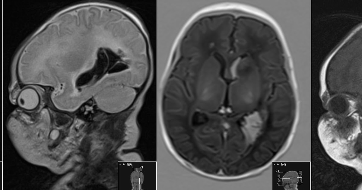
Radiology MRI: Neonatal Intraventricular Hemorrhage

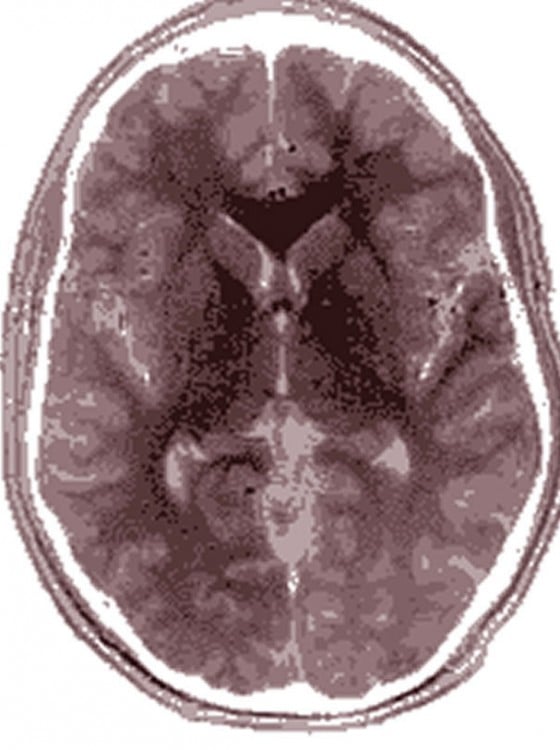
Radiodiagnosis - Imaging is Amazing-Interesting cases: Phenytoin associated Cerebellar atrophy - MRI
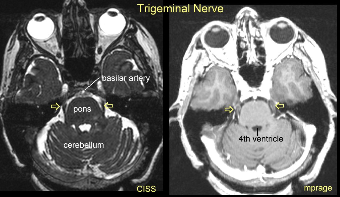
Gamma Knife for Trigeminal Neuralgia

Radiology MRI: Occipital Infarct

Mouse brain seen in sharpest detail ever | Kurzweil

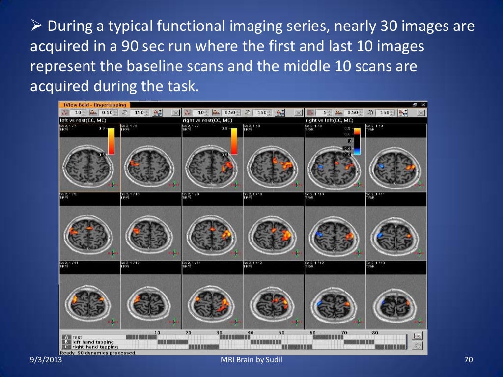
UMD PSYC E-News: RA positions focusing on functional MRI and Autism Spectrum Disorder!

February | 2011 | Thought Broadcast
MRI Procedure of Brain
Brain Anatomy Differences Between Autistic and Typically Developing Individuals Are ...

Neuroanatomy - encyclopedia article - Citizendium

Gamma Knife for Trigeminal Neuralgia
A Hybrid Approach for Automatic Classification of Brain MRI Using Genetic Algorithm and Support ...

MRI Brain Planning

Medical Legal Demonstrative Evidence
MRI SECTIONAL ANATOMY OF BRAIN

MRI of Brain - Stock Image - M134/0816 - Science Photo Library

MRI Brain Poster | Zazzle.com
Post a Comment for "40 brain mri with labels"